Lymphoid Cells

CD10
CD10 – Cluster of Differentiation 10
CALLA – Common Acute Lymphocytic Leukemia Antigen
MME – Membrane Metallo-Endopeptidase
NEP – Neutral Endopeptidase
SFE
A type II transmembrane protein found on pre-B cells, germinal-center B cells, some neutrophils, kidney cells, T-cell precursors, and epithelial cells that acts as a zinc metalloprotease cleaving peptide bonds on the amino side of hydrophobic amino acids; expressed in acute lymphocytic leukemia and follicular-center-cell lymphomas. Cell membrane metallopeptidase widely distributed in hematopoietic cells and their neoplasms. Important in diagnosis of preB-AL.
Reactivity: Follicle center cells, Follicular Lymphoma (FL), some Diffuse Large B-cell Lymphoma (DLBCL), Acute B Lymphoblastic Leukemia (pre-B-ALL), Acute T Lymphoblastic Leukemia/Lymphoma (pre-T-ALL), thymocytes, Burkitt’s Lymphoma (BL).
Pathology Information for CD10
CD-10 Receptor:
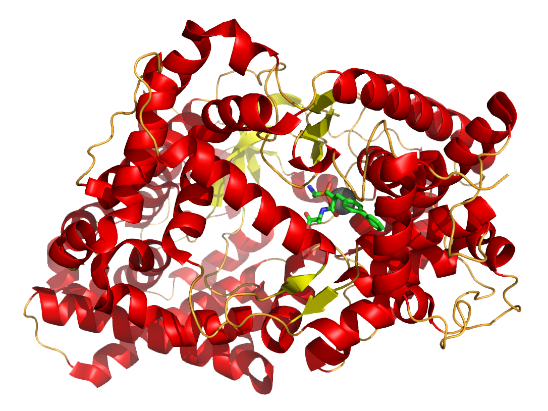
The structure of neprilysin, a zinc metalloprotease involved in the metabolism of extracellular regulatory peptides such as the enkephalins and substance P. Neprilysin in neural tissue is the major protease degrading amyloid beta, a peptide fragment whose abnormal misfolding and aggregation is associated with Alzheimer’s disease. The gray sphere represents a zinc atom; the green structure is a zinc-chelating enzyme inhibitor. (Opabinia regalis. Self-created from PDB ID 1R1H using PyMol)
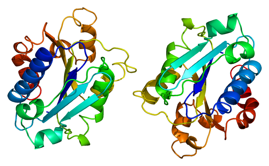
CD5
CD5 – Cluster of Differentiation 5
LEU1
T1
CD5 is a cluster of differentiation expressed on the surface of T cells and in a subset of murine B cells known as B-1a (Important in identification of T cells and most T cell lymphomas, some low grade B cell lymphomas, thymic carcinoma). CD5 is type I transmembrane protein found on T cells, thymocytes, and some B cells that is a ligand for CD72 and is involved in cellular activation or adhesion; expressed in B-cell chronic lymphocytic leukemia and T-cell lymphoma.
Reactivity: T cells, Chronic Lymphocytic Leukemia/Small Cell Lymphoma (B-CLL/SLL), Mantle Cell Lymphoma (MCL).
Pathology Information for CD5
CD-5 Receptor:
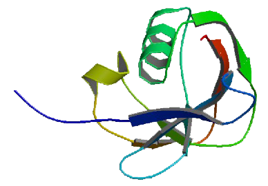
Structure of CD5 protein
CD200
CD200 – Cluster of Differentiation 200
OX-2 Membrane Glycoprotein
MOX1
MOX2
MRC
CD200 is a protein encoded by the CD200 gene. An immunoadhesin that may deliver immunosuppressive signals and regulate autoimmune disorders. Inhibitory for macrophage lineage cells. CD200 is a type-1 membrane glycoprotein belonging to the immunoglobulin superfamily. May regulate myeloid cell activity and delivers an inhibitory signal for the macrophage lineage in diverse tissues. CD200 is overexpressed by many different types of hematological and solid tumors. Biotech company Trillium has developed an investigative anti-CD200 monoclonal antibody.
Reactivity: CD200 has an important role in the differential diagnosis of mature B‐cell neoplasms. CD200-CD200R Interaction: An Important Regulator After Stroke.
Pathology Information for CD200
CD34
CD34 – Cluster of Differentiation 34
Hematopoietic Progenitor Cell Antigen CD34
CD34 is a transmembrane phosphoglycoprotein protein encoded by the CD34 gene. CD34 is a stem cell marker, adhesion, found on hematopoietic precursors (found in high concentrations in umbilical cord blood), capillary endothelium, and embryonic fibroblasts.
Reactivity: Myeloblasts, Lymphoblasts, Endothelial cells
Pathology Information for CD34
CD38
CD38 – Cluster of Differentiation 38
Cyclic ADP Ribose Hydrolase
ADPRC1
CD38 is a glycoprotein found on the surface of many immune cells (white blood cells), including CD4, CD8, B lymphocytes and natural killer cells. CD38 also functions in cell adhesion, signal transduction and calcium signaling. CD38 is a marker of cellular activation expressed by plasma cells, T cells, NK cells and other hematopoietic cell types during various stages of maturation. CD 38 is involved in ecto-ADP-ribosyl cyclase and cell activation on many hematopoietic, plasma, and B & T activated cells; marker increases with HIV seroconversion, coexpression with CD8 associated with progression (indicates persistent viral stimulation). Some antibodies targeting CD38 are being tested in multiple myeloma and non-Hodgkin’s lymphoma e.g. Daratumumab or Celgene’s MOR202.
Reactivity: Plasma Cells, Activated T and B Cells, Subset Chronic Lymphocytic Leukemia/Small Cell Lymphoma (B-CLL/SLL), Epithelial Cells.
Pathology Information for CD38
CD-38 Receptor:
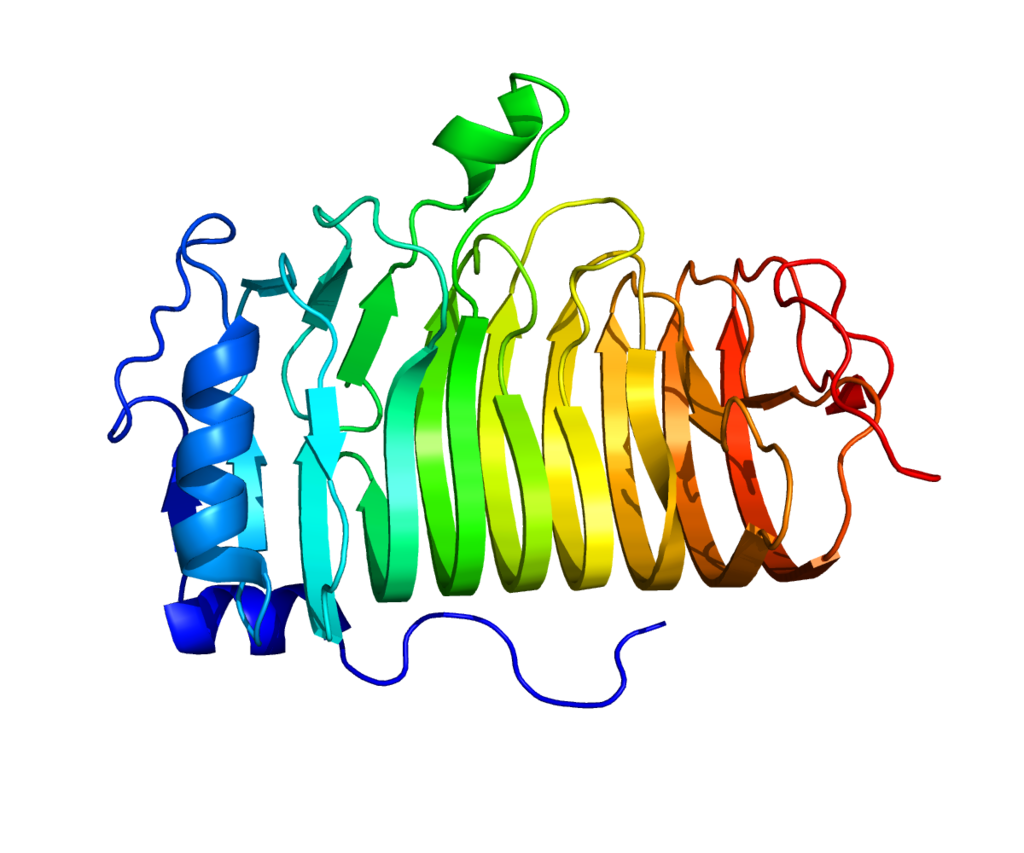
Structure of the CD38 protein. Based on PyMOL rendering of PDB 1yh3.
CD20
CD20 – Cluster of Differentiation 20
B-lymphocyte antigen CD20
CVID5, LEU-16, MS4A2, S7, L26
CD20 is a type III transmembrane protein found on B cells that forms a calcium channel in the cell membrane allowing for the influx of calcium required for cell activation; expressed in B-cell lymphomas, hairy cell leukemia, and B-cell chronic lymphocytic leukemia. Important for therapy of those diseases, as antibodies against CD20 exist: e.g. Rituximab and Ofatumumab, with several more in development. Similarly, anti-CD20 monoclonal antibody Ocrelizumab is in trials for multiple sclerosis.
Reactivity: B Cells, Rare Plasma Cell Myelomas.
Pathology Information for CD20
CD-20 Receptor:
Structure of protein MS4A1.Based on PyMOL rendering of PDB 1S8B.
CD19
CD19 – Cluster of Differentiation 19
B-lymphocyte antigen CD19
B-Lymphocyte Surface Antigen B4
T-Cell Surface Antigen Leu-12
CVID3
CD19 is a transmembrane protein that is encoded by the gene CD19. Common B cell marker. Expressed by B cells and follicular dendritic cells. Early B lineage-restricted antigen present on most pre-B cells, B-ALL and B-CLL cells. CD19 is B-lymphocyte surface antigen B4, component of the B-cell co-receptor; highly represented in B-cell malignancies, CD19 is the target of several CAR-T and mAb cancer drugs in development e.g. Juno JCAR015, Kite KTE-C19 CAR, Novartis CTL019, Morphosys MOR208, Macrogenics MGD011, Affimed AFM11.
Reactivity: B Cells, Acute B Lymphoblastic Leukemia (Pre B-ALL), Subset of Acute Myelogenous Leukemia (AML – AML1/ETO with t(8;21)).
Pathology Information for CD19
CD45
CD45 – Cluster of Differentiation 45
PTPRC – Protein Tyrosine Phosphatase, Receptor Type
B220, GP180, CD45R, LY5, T200
LCA – Leukocyte Common Antigen
CD45 is an enzyme that is encoded by the PTPRC gene. CD45 is commonly used marker of hematopoietic cells except erythrocytes and platelets; plays a major role in immune system. CD45 is a leucocyte common antigen, a type I transmembrane protein present on all hemopoietic cells except erythrocytes that assists in cell activation; expressed in lymphomas, B-cell chronic lymphocytic leukemia, hairy cell leukemia, and acute nonlymphocytic leukemia.
Reactivity: CD45 is a good indicator of the hematopoietic nature of tumors. May assist in classification of lymphomas and leukemias. Confirm presence of inflammatory cells, including intestinal intraepithelial lymphocytes. CD45 and keratin expression are mutually exclusive in diagnostic pathology (keratin reactivity is common of abnormal epithelial differentiation, which result in disturbed barrier function of human skin (carcinomas and some sarcomas)).
Pathology Information for CD45
CD-45 Receptor:
Structure of the PTPRC protein. Based on PyMOL rendering of PDB 1ygr. .

CD4
CD4 – Cluster of Differentiation 4
It was discovered in the late 1970s and was originally known as LEU-3 and T4 (after the OKT4 monoclonal antibody that reacted with it) before being named CD4 in 1984. The CD4 protein is encoded by the CD4 gene. CD4 is a marker of T helper cells (used to classify lymphomas and inflammatory conditions), important in T cell activation and receptor for HIV (serum levels are marker of HIV disease progression and response to HAART therapy (CD4+ cells are killed by HIV)). Serum levels used to diagnose idiopathic CD4 lymphocytopenia. CD4 is a co-receptor for MHC Class II (with TCR, T-cell receptor); found on the surface of immune cells such as T helper cells, monocytes, macrophages, and dendritic cells. Used by HIV to enter T cells: in HIV infection. CD8+ cytotoxic T cells recognize and kill infected cells hence they are predominantly for protection against intracellular pathogens, e.g. viruses, and some bacteria, i.e. Rickettsiae.
Reactivity: T Cells (helper/inducer), Monocytes, Myeloblasts, NK cell lymphoma.
Pathology Information for CD4
CD2
CD2 – Cluster of Differentiation 2
T-cell Surface Antigen T11/LEU-5
LFA-2,
LFA-3 Receptor
Erythrocyte Receptor
Rosette Receptor
CD2 is a cell adhesion molecule found on the surface of T cells and natural killer (NK) cells. Early T cell marker expressed on all peripheral blood T cells, 95% of thymocytes, NK cells, but no B cells. Gene is at 1p13.1; protein is member of immunoglobulin superfamily. CD 2 is a type I transmembrane protein found on thymocytes, T cells, and some natural killer cells that acts as a ligand for CD58 and CD59 and is involved in signal transduction and cell adhesion; expressed in T-cell acute lymphoblastic leukemia and T-cell lymphoma.
Reactivity: T Cells, Large Granular Lymphocytes (LGL), NK Cells, Acute Promyelocytic Leukemia (APL), Neoplastic Mast Cells.
CD-2 Receptor:
Extracytoplasmic segment of the Uman CD2 antigen, in green the protein, at the upper left the adesion domain, where is clearly recognizable the Ig structure with the three loops. There are also shown the N-ac-GLU residues where is developed a poliantennary glication based on D-man and Glu. On the right side is the proximal ig domain. .
- Early T cell marker expressed on all peripheral blood T cells, 95% of thymocytes, NK cells but no B cells (OMIM: 186990 [Accessed 13 May 2021], J Biol Chem 2010;285:41755)
- Gene is at 1p13.1; protein is member of immunoglobulin superfamily
- Also called E rosette receptor because CD2 antibodies inhibit formation of rosettes with sheep erythrocytes
- Also called LFA2 (leukocyte function antigen), T11
- Pathophysiology:
- Binds CD58 (LFA3) on antigen presenting cells, which enables T cells to respond to lower concentrations of antigen (J Exp Med 1999;190:1383)
- Mediates adhesion between T cells and antigen presenting cells, induces costimulatory signals in T cells and T cell cytokine production, inhibits apoptosis of activated peripheral T cells (Immunology 2013;139:48)
- Regulates T cell anergy
- Regulates T and NK mediated cytolysis
- CD2 distinguishes 2 subsets of human plasmacytoid dendritic cells with distinct phenotype and functions (J Immunol 2009;182:6815)
- CD2 (low) plasmacytoid dendritic cells are depleted in HIV+ patients (Cell Mol Immunol 2011;8:441)
- CD2 promotes NK cell membrane nanotube formation (PLoS One 2012;7:e47664)
- Clinical features:
- Abnormal levels of CD2 expression by flow cytometry are frequently observed in mature T cell malignancies but clinical significance is unclear because of variability of CD2 expression in normal T cells (Cytometry B Clin Cytom 2010;78:169)
- Rare aberrant expression in B cell lymphomas (Appl Immunohistochem Mol Morphol 2011;19:579)
- High CD2 expression identifies latently infected resting memory CD4(+) T cells in vivo (J Virol 2013;87:9148)
- Siplizumab, an anti CD2 monoclonal antibody, was previously studied to treat plaque psoriasis; no current clinical trials as of last update (Int J Dermatol 2010;49:818)
- Uses by pathologists: T cell marker (membranous staining), although CD3 is more commonly used; marker of systemic mastocytosis
- Positive staining – normal:
- Thymocytes (95%), mature peripheral T cells (almost all), NK cells (80 – 90%), mast cells
- Variable thymic B cells (50%), Langerhans cells
- Positive staining – disease:
- T-ALL, other T cell lymphoma / leukemia, acute promyelocytic leukemia microgranular variant / FAB M3v (Leukemia 1995;9:1461)
- AML with pseudo-Chédiak-Higashi anomaly (Am J Clin Pathol 2006;125:791)
- Systemic mastocytosis (a minor criteria, Hum Pathol 2001;32:545)
- And other mast cell disease, thymoma (lymphocyte predominant)
- Variable:
- Acute myeloid lymphoma M0
- Rare:
- Pyothorax associated B cell lymphoma (Am J Surg Pathol 2002;26:724)
- Other non-Hodgkin B cell lymphomas, Reed-Sternberg / Hodgkins cells (Mod Pathol 2005;18:1542)
- Myeloma (Mod Pathol 1990;3:302)
- Myeloid leukemia
- Negative staining: B cells, basophils, nonhematopoietic neoplasms and mast cells in nonmastocytosis disorders (Am J Clin Pathol 2003;120:64)
CD56
CD56 – Cluster of Differentiation 56
NCAM – Neural cell adhesion molecule
CD56 is a homophilic binding glycoprotein (with role in cell–cell adhesion) expressed on the surface of neurons, glia and skeletal muscle. CD 56 is a 140 kD isoform of NCAM (neural cell adhesion molecule), a marker for natural killer cells and some T-lymphocytes.
Reactivity: NK cells, T-Cell Large Granular Leukemia (LGL).
Pathology Information for CD56
CD5
CD5 – Cluster of Differentiation 5
LEU1
T1
CD5 is a cluster of differentiation expressed on the surface of T cells and in a subset of murine B cells known as B-1a (Important in identification of T cells and most T cell lymphomas, some low grade B cell lymphomas, thymic carcinoma). CD5 is type I transmembrane protein found on T cells, thymocytes, and some B cells that is a ligand for CD72 and is involved in cellular activation or adhesion; expressed in B-cell chronic lymphocytic leukemia and T-cell lymphoma.
Reactivity: T cells, Chronic lymphocytic leukemia/small cell lymphoma (B-CLL/SLL), Mantle Cell Lymphoma (MCL).
Pathology Information for CD5
CD5 Receptor:
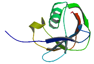
Structure of CD5 protein
CD34
CD34 – Cluster of Differentiation 34
Hematopoietic Progenitor Cell Antigen CD34
CD34 is a transmembrane phosphoglycoprotein protein encoded by the CD34 gene. CD34 is a stem cell marker, adhesion, found on hematopoietic precursors (found in high concentrations in umbilical cord blood), capillary endothelium, and embryonic fibroblasts.
Reactivity: Myeloblasts, Lymphoblasts, Endothelial cells
Pathology Information for CD34
CD3
CD3 – Cluster of Differentiation 3
OKT3
CD3 is a protein complex and T cell co-receptor that is involved in activating both the cytotoxic T cell (CD8+ naive T cells) and T helper cells (CD4+ naive T cells). Member of immunoglobulin superfamily on 11q23. Common antibody for identifying T cells (the signaling component of the T cell receptor (TCR) complex).
Reactivity: T Cells, Primary Effusion Lymphoma.
Pathology Information for CD3
The T-Cell Receptor Complex:
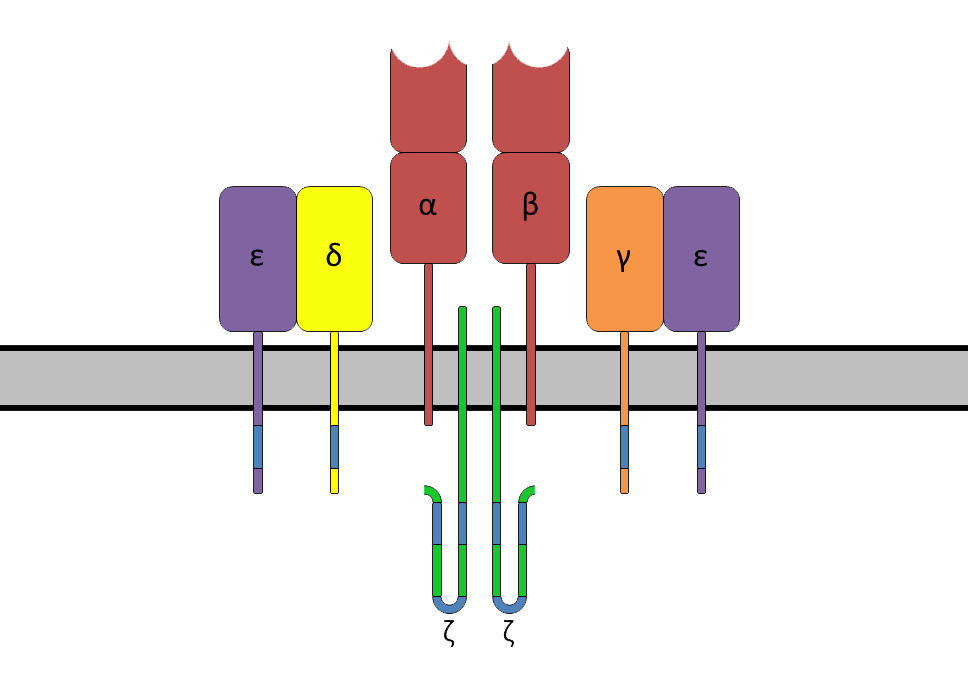
The T cell receptor complex with TCR-α and TCR-β chains (top), ζ-chain (CD247) accessory molecules (bottom) and CD3 (represented by CD3γ, CD3δ and two CD3ε).
CD8
CD8 – Cluster of Differentiation 8
OKT8
CD8 is a transmembrane glycoprotein that serves as a co-receptor for the T-cell receptor (TCR). CD8 is a cytotoxic T cell marker. CD8+ cells are called T cell suppressor cells, cytotoxic T cells. Along with the TCR, the CD8 co-receptor plays a role in T cell signaling and aiding with cytotoxic T cell antigen interactions. CD8 binds to a major histocompatibility complex (MHC) molecule, but is specific for the MHC class I protein. CD8 is a co-receptor (with TCR, T-cell receptor) for MHC Class I; mostly found on cytotoxic T cells, but also on natural killer cells, cortical thymocytes, and a subset of myeloid dendritic cells. In HIV infection, CD8+ cytotoxic T cells recognise and kill infected CD4+ helper T cells, which are critical for the body’s immunity. In HBV infection CD8+ cytotoxic T cells are involved in liver injury by killing infected cells and by producing antiviral cytokines capable of purging HBV from viable hepatocytes. There are two isoforms of the protein, alpha and beta, each encoded by a different gene.
Reactivity: T Cells (Suppressor/Cytotoxic), Large Granular Lymphocytes T-Cell Large Granular Leukemia (LGL).
Pathology Information for CD8
CD-8 Receptor:
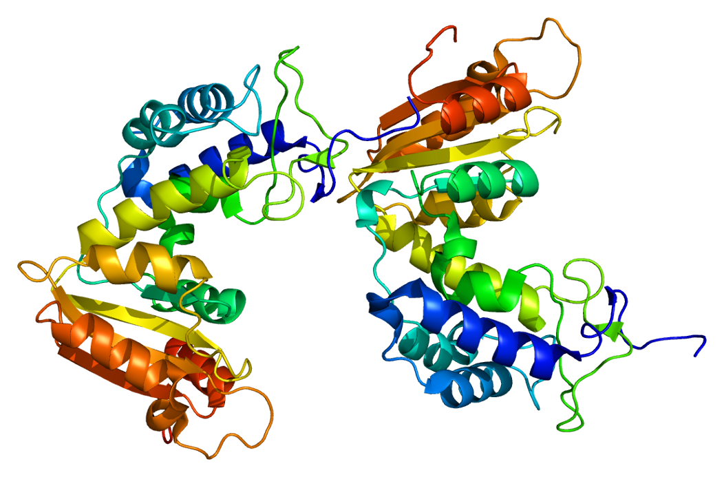
Schematic representation of the heterodimeric CD8 co-receptor. A schematical representation of CD8-receptor, found on cytotoxic T cells. It consists of an α and a β chain and a carboxy terminus (C) in the cell-membrane.
CD7
CD7 – Cluster of Differentiation 7
GP40, LEU-9, TP41, Tp40
CD7 is T cell cell surface protein (encoded by the CD7 gene) that plays important role in T cell-B cell interaction in early lymphoid development. CD7 is a member of immunoglobulin superfamily at 17q25.2-25.3. Membrane expression early during T cell development, before TCR rearrangement; persists until terminal stages of T cell development. CD7 has costimulatory activity for T cells. Binds galectin-1 and is essential for galectin-1-mediated T-cell death; loss of CD7 expression and altered cellular glycosylation may contribute to apoptosis resistance in mycosis fungoides. CD7 is a type I transmembrane protein found on thymocytes, some T cells, monocytes, natural killer cells, and hematopoietic stem cells; expressed in patients with mycosis fungoides, some patients with T-cell acute lymphoblastic leukemia, and a few patients with acute nonlymphocytic leukemia.
Reactivity: T Cells, Some Myeloblasts.
Pathology Information for CD7
CD45
CD45 – Cluster of Differentiation 45
PTPRC – Protein Tyrosine Phosphatase, Receptor Type
B220, GP180, CD45R, LY5, T200
LCA – Leukocyte Common Antigen
CD45 is an enzyme that is encoded by the PTPRC gene. CD45 is commonly used marker of hematopoietic cells except erythrocytes and platelets; plays a major role in immune system. CD45 is a leucocyte common antigen, a type I transmembrane protein present on all hemopoietic cells except erythrocytes that assists in cell activation; expressed in lymphomas, B-cell chronic lymphocytic leukemia, hairy cell leukemia, and acute nonlymphocytic leukemia.
Reactivity: CD45 is a good indicator of the hematopoietic nature of tumors. May assist in classification of lymphomas and leukemias. Confirm presence of inflammatory cells, including intestinal intraepithelial lymphocytes. CD45 and keratin expression are mutually exclusive in diagnostic pathology (keratin reactivity is common of abnormal epithelial differentiation, which result in disturbed barrier function of human skin (carcinomas and some sarcomas)).
Pathology Information for CD45
CD-45 Receptor:
Structure of the PTPRC protein. Based on PyMOL rendering of PDB 1ygr. .
Myeloid Cells

CD16
CD16 – Cluster of Differentiation 16
FcγRIII
Exists in two different forms: FcγRIIIa (CD16a) and FcγRIIIb (CD16b)
CD16 is a cluster of differentiation molecule found on the surface of natural killer cells, neutrophils, monocytes, and macrophages. FcγRIII, a low-affinity Fc receptor for IgG. Found on NK cells, macrophages, and neutrophils. Cd16 also known as Fc gamma receptor III A, FCGR3A, low affinity immunoglobulin gamma Fc region receptor III-A. CD14+ CD16+ monocytes have increased capacity to produce proinflammatory cytokines such as TNF-alpha, and are elevated in various inflammatory diseases, including coronary. CD16a polymorphisms influence: (a) the severity but not the incidence of IgA nephropathy in Japanese patientsartery disease, (b) pathogenesis of coronary artery disease, (c) clinical response to rituximab, and infliximab for Crohn disease. Loss of CD16a in children is associated with recurrent viral infections.
Reactivity: Granulocytes, NK cells, T-Cell Large Granular Leukemia (LGL).
Pathology Information for CD16
CD7
CD7 – Cluster of Differentiation 7
GP40, LEU-9, TP41, Tp40
CD7 is T cell cell surface protein (encoded by the CD7 gene) that plays important role in T cell-B cell interaction in early lymphoid development. CD7 is a member of immunoglobulin superfamily at 17q25.2-25.3. Membrane expression early during T cell development, before TCR rearrangement; persists until terminal stages of T cell development. CD7 has costimulatory activity for T cells. Binds galectin-1 and is essential for galectin-1-mediated T-cell death; loss of CD7 expression and altered cellular glycosylation may contribute to apoptosis resistance in mycosis fungoides. CD7 is a type I transmembrane protein found on thymocytes, some T cells, monocytes, natural killer cells, and hematopoietic stem cells; expressed in patients with mycosis fungoides, some patients with T-cell acute lymphoblastic leukemia, and a few patients with acute nonlymphocytic leukemia.
Reactivity: T Cells, Some Myeloblasts.
Pathology Information for CD7
CD10
CD10 – Cluster of Differentiation 10
CALLA – Common Acute Lymphocytic Leukemia Antigen
MME – Membrane Metallo-Endopeptidase
NEP – Neutral Endopeptidase
SFE
A type II transmembrane protein found on pre-B cells, germinal-center B cells, some neutrophils, kidney cells, T-cell precursors, and epithelial cells that acts as a zinc metalloprotease cleaving peptide bonds on the amino side of hydrophobic amino acids; expressed in acute lymphocytic leukemia and follicular-center-cell lymphomas. Cell membrane metallopeptidase widely distributed in hematopoietic cells and their neoplasms. Important in diagnosis of preB-AL.
Reactivity: Follicle center cells, Follicular Lymphoma (FL), some Diffuse Large B-cell Lymphoma (DLBCL), Acute B Lymphoblastic Leukemia (pre-B-ALL), Acute T Lymphoblastic Leukemia/Lymphoma (pre-T-ALL), thymocytes, Burkitt’s Lymphoma (BL).
Pathology Information for CD10
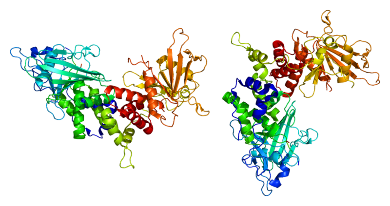
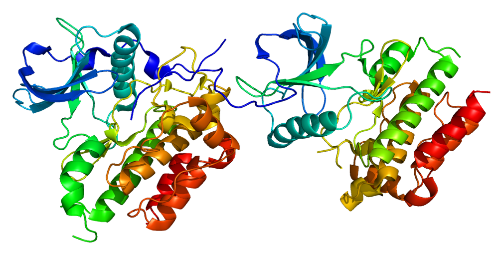
CD-10 Receptor:
The structure of neprilysin, a zinc metalloprotease involved in the metabolism of extracellular regulatory peptides such as the enkephalins and substance P. Neprilysin in neural tissue is the major protease degrading amyloid beta, a peptide fragment whose abnormal misfolding and aggregation is associated with Alzheimer’s disease. The gray sphere represents a zinc atom; the green structure is a zinc-chelating enzyme inhibitor. (Opabinia regalis. Self-created from PDB ID 1R1H using PyMol)
CD13
CD13 – Cluster of Differentiation 13
Membrane Alanyl Aminopeptidase
APN – Aminopeptidase N
GP150, LAP1, P150, PEPN
CD13 is an enzyme that is encoded by the ANPEP gene. CD13 is a myeloid antigen (although CD33 is more specific). CD 13 is a zinc metalloproteinase, also known as aminopeptidase N, which is found naturally on myelomonocytic cells from early differentiation through maturity; usually present on acute myeloid leukemia blasts and rarely found in some forms of lymphoma and lymphocytic leukemia.
Reactivity: Myeloid Cells, Rare Acute B Lymphoblastic Leukemia (pre-B-ALL).
Pathology Information for CD13
CD64
CD64 – Cluster of Differentiation 64
FcγR – Fc-gamma receptor 1
FCGR1A, FCGR1B,FCGR1C
CD64 is a type of integral membrane glycoprotein known as an Fc receptor that binds monomeric IgG-type antibodies with high affinity. After binding IgG, CD64 interacts with an accessory chain known as the common γ chain (γ chain), which possesses an ITAM motif that is necessary for triggering cellular activation. Important in phagocytosis via receptor-mediated endocytosis of IgG-antigen complexes. Mediates antigen capture for presentation to T cells, antibody-dependent cellular cytotoxicity, release of cytokines and reactive oxygen intermediates. Binds to C reaction protein and mediates its effects. Denge fever immune complexes enter macrophages via CD64. CD64 is found on macrophages and monocytes.
Reactivity: High neutrophil CD64 levels predict sepsis, early onset of infection in neonates and infection in rheumatoid arthritis patients.
Pathology Information for CD64
CD34
CD34 – Cluster of Differentiation 34
Hematopoietic Progenitor Cell Antigen CD34
CD34 is a transmembrane phosphoglycoprotein protein encoded by the CD34 gene. CD34 is a stem cell marker, adhesion, found on hematopoietic precursors (found in high concentrations in umbilical cord blood), capillary endothelium, and embryonic fibroblasts.
Reactivity: Myeloblasts, Lymphoblasts, Endothelial cells
Pathology Information for CD34
CD14
CD14 – Cluster of Differentiation 14
Lipopolysaccharide (LPS) Receptor
Monocyte Differentiation Antigen
CD14 is a protein made mostly by macrophages as part of the innate immune system. It helps to detect bacteria in the body by binding lipopolysaccharide (LPS), a pathogen-associated molecular pattern (PAMP). CD14 is a pattern recognition receptor that detects antigenic molecules on the surface of various microorganisms. Mutations can prevent adequate inflammatory response to infection, leading to systemic infections. CD 14 is a membrane protein found on macrophages which binds to bacterial lipopolysaccharide.
Reactivity: Monocytes
Pathology Information for CD14
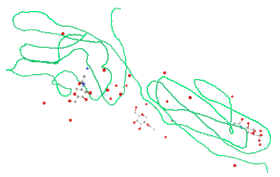
CD-14 Receptor:
Crystal structure of CD14 and its implications for lipopolysaccharide signaling, illustrated by Jmol Version 12.0.41. .
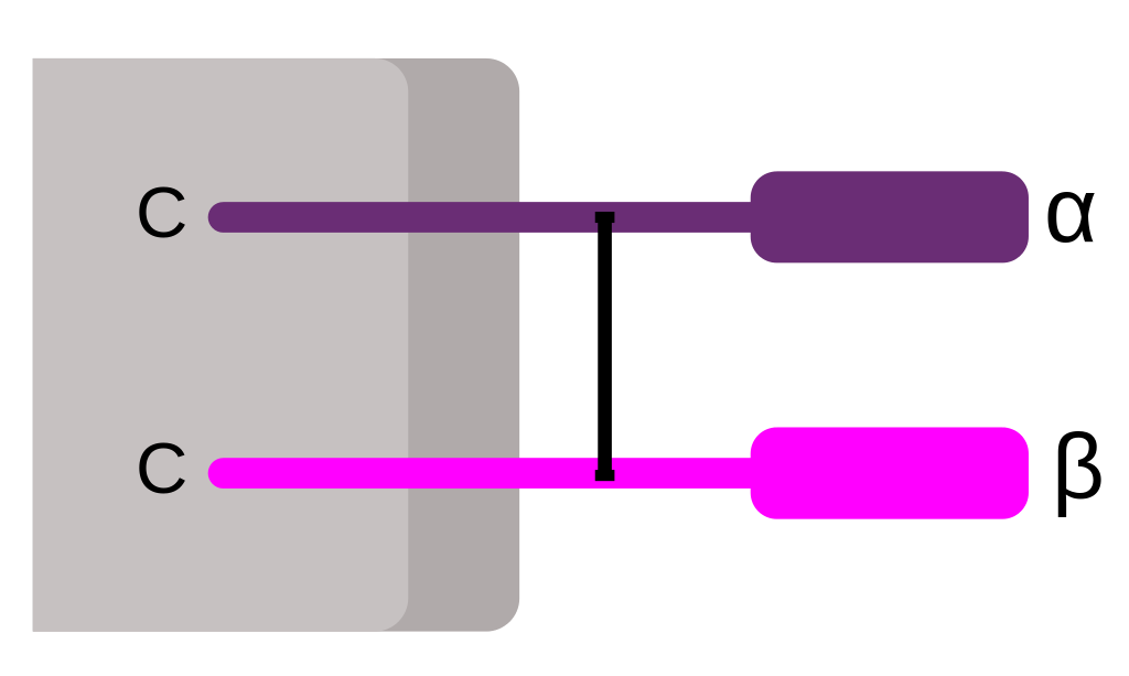
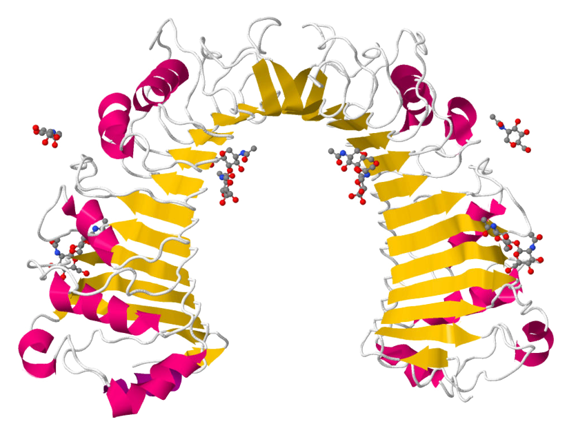
HLA-DR
HLA-DR – Human Leukocyte Antigen – DR Isotype
MHC class II, DR
HLA-DR (Ia)
HLA-DR is an MHC (major histocompatibility complex) class II cell surface receptor encoded by the human leukocyte antigen complex on chromosome 6 region 6p21.31. The complex of HLA-DR and peptide, generally between 9 and 30 amino acids in length, constitutes a ligand for the T-cell receptor (TCR). HLA (human leukocyte antigens) were originally defined as cell surface antigens that mediate graft-versus-host disease. Identification of these antigens has led to greater success and longevity in organ transplant. Antigen presenting cells use HLA-DR molecules to present protein fragments (processed antigen) to T cells, a key event in induction of T cell responses.
Reactivity: Acute Myelogenous Leukemia (AML), except APL, B Cells Monocytes.
Pathology Information for HLA-DR
DR Receptor:
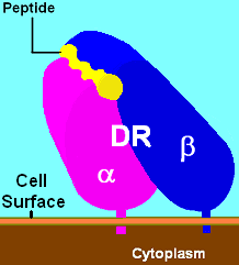
Illustration of DR with bound ligand (yellow).
CD11b
CD11b – Cluster of Differentiation Molecule 11B
ITGAM, CR3A, MAC-1, MAC1A, MO1A, SLEB6, Integrin Subunit Alpha M
Integrin Alpha M (ITGAM); the alpha subunit of Mac-1 (Macrophage-1 antigen), the CR3 complement receptor which consists of CD11b and CD18. CR3 is a human cell surface receptor, found on polymorphonuclear leukocytes (mostly neutrophils), NK cells, and mononuclear phagocytes like macrophages, which is capable of recognizing and binding to many molecules found on the surfaces of invading bacteria. Binding to the receptor causes phagocytosis and destruction of the foreign cell. Alpha component of an integrin at 16p11-13.1 that mediates adhesion.
Reactivity: Granulocytes, Monocytes.
Pathology Information for CDCD11b
CD11b Receptor:
Structure of the ITGAM protein. Based on PyMOL rendering of PDB 1bho. .
CD45
CD45 – Cluster of Differentiation 45
PTPRC – Protein Tyrosine Phosphatase, Receptor Type
B220, GP180, CD45R, LY5, T200
LCA – Leukocyte Common Antigen
CD45 is an enzyme that is encoded by the PTPRC gene. CD45 is commonly used marker of hematopoietic cells except erythrocytes and platelets; plays a major role in immune system. CD45 is a leucocyte common antigen, a type I transmembrane protein present on all hemopoietic cells except erythrocytes that assists in cell activation; expressed in lymphomas, B-cell chronic lymphocytic leukemia, hairy cell leukemia, and acute nonlymphocytic leukemia.
Reactivity: CD45 is a good indicator of the hematopoietic nature of tumors. May assist in classification of lymphomas and leukemias. Confirm presence of inflammatory cells, including intestinal intraepithelial lymphocytes. CD45 and keratin expression are mutually exclusive in diagnostic pathology (keratin reactivity is common of abnormal epithelial differentiation, which result in disturbed barrier function of human skin (carcinomas and some sarcomas)).
Pathology Information for CD45
CD-45 Receptor:
Structure of the PTPRC protein. Based on PyMOL rendering of PDB 1ygr. .

CD15
CD15 – Cluster of Differentiation 15
LeuM1, Lewis X, 3-Fucosyl-N-Acetyl-Lactosamine
CD15 is a tetrasaccharide carbohydrate which is usually attached to O-glycans on the surface of cells. It is known to play a vital role in cell-to-cell recognition processes. CD15 is a carbohydrate adhesion molecule (not a protein) that mediates phagocytosis and chemotaxis, found on neutrophils; expressed in patients with Hodgkin disease, some B-cell chronic lymphocytic leukemias, acute lymphoblastic leukemias, and most acute nonlymphocytic leukemias. It is also called Lewis x and SSEA-1 (stage specific embryonic antigen 1) and represents a marker for murine pluripotent stem cells, in which it plays an important role in adhesion and migration of the cells in the preimplantation embryo.
Reactivity: Granulocytes, Hodgkin’s Lymphoma.
Pathology Information for CD15
CD123
CD123 – Cluster of Differentiation 123
IL-3R – Interleukin-3 Receptor
CD123 is a molecule found on cells which helps transmit the signal of interleukin-3, a soluble cytokine important in the immune system. It is found on pluripotent progenitor cells, induces tyrosine phosphorylation within the cell, and promotes proliferation and differentiation within the hematopoietic cell lines. Associates with the GM-CSF receptor (GMR beta).
Reactivity: Expressed in Hematologic Malignancies. Reacts with plasmacytoid dendritic cells in situ.
Pathology Information for CD123
CD117
CD117 – Cluster of Differentiation 117
SCFR – Mast/Stem Cell Growth Factor Receptor
Proto-Oncogene C-KIT
KIT, PBT,
KIT Proto-Oncogene, Receptor Tyrosine Kinase,
MASTC
CD117 is a c-kit, the receptor for Stem Cell Factor, a glycoprotein that regulates cellular differentiation, particularly in hematopoiesis. CD117 is a proto-oncogene activated in GIST tumors. Receptor for kit protein, a 145 kD tyrosine kinase growth factor receptor protein important for development and survival of mast cells, hematopoietic stem cells, melanocytes, germ cells, interstitial cells of Cajal. Has activating or gain of function mutations in most GIST tumors, often at exon 11, less often at exons 9 and 13. Tyrosine kinase activity of c-kit in GIST and bcl-abl overexpression in CML are inhibited by imatinib mesylate, a tyrosine kinase inhibitor used to treat these diseases.
Reactivity: Acute Myelogenous Leukemia (AML), Mast Cells, Stromal Tumors (GIST), Plasma Cells.
Pathology Information for CD117
CD-117 Receptor:
Structure of the KIT protein. Based on PyMOL rendering of PDB 1pkg. .
CD13
CD13 – Cluster of Differentiation 13
Membrane Alanyl Aminopeptidase
APN – Aminopeptidase N
GP150, LAP1, P150, PEPN
CD13 is an enzyme that is encoded by the ANPEP gene. CD13 is a myeloid antigen (although CD33 is more specific). CD 13 is a zinc metalloproteinase, also known as aminopeptidase N, which is found naturally on myelomonocytic cells from early differentiation through maturity; usually present on acute myeloid leukemia blasts and rarely found in some forms of lymphoma and lymphocytic leukemia.
Reactivity: Myeloid Cells, Rare Acute B Lymphoblastic Leukemia (pre-B-ALL).
Pathology Information for CD13
CD33
CD33 – Cluster of Differentiation 33
CD33 is a marker of unknown function found on immature myeloid cells, including acute myeloid leukemia blasts and mature monocytes. Anti-CD33 monoclonal antibodies are extensively used for the diagnosis of all types of AMLs. Classic myeloid antigen.
Reactivity: Myeloid Cells, Rare Pre B-ALL (Acute B Lymphoblastic Leukemia), Rare Blastic NK Lymphoma.
Pathology Information for CD33
CD34
CD34 – Cluster of Differentiation 34
Hematopoietic Progenitor Cell Antigen CD34
CD34 is a transmembrane phosphoglycoprotein protein encoded by the CD34 gene. CD34 is a stem cell marker, adhesion, found on hematopoietic precursors (found in high concentrations in umbilical cord blood), capillary endothelium, and embryonic fibroblasts.
Reactivity: Myeloblasts, Lymphoblasts, Endothelial cells
Pathology Information for CD34
CD38
CD38 – Cluster of Differentiation 38
Cyclic ADP Ribose Hydrolase
ADPRC1
CD38 is a glycoprotein found on the surface of many immune cells (white blood cells), including CD4, CD8, B lymphocytes and natural killer cells. CD38 also functions in cell adhesion, signal transduction and calcium signaling. CD38 is a marker of cellular activation expressed by plasma cells, T cells, NK cells and other hematopoietic cell types during various stages of maturation. CD 38 is involved in ecto-ADP-ribosyl cyclase and cell activation on many hematopoietic, plasma, and B & T activated cells; marker increases with HIV seroconversion, coexpression with CD8 associated with progression (indicates persistent viral stimulation). Some antibodies targeting CD38 are being tested in multiple myeloma and non-Hodgkin’s lymphoma e.g. Daratumumab or Celgene’s MOR202.
Reactivity: Plasma Cells, Activated T and B Cells, Subset Chronic Lymphocytic Leukemia/Small Cell Lymphoma (B-CLL/SLL), Epithelial Cells.
Pathology Information for CD38
CD-38 Receptor:
Structure of the CD38 protein. Based on PyMOL rendering of PDB 1yh3.
HLA-DR
HLA-DR – Human Leukocyte Antigen – DR Isotype
MHC class II, DR
HLA-DR (Ia)
HLA-DR is an MHC (major histocompatibility complex) class II cell surface receptor encoded by the human leukocyte antigen complex on chromosome 6 region 6p21.31. The complex of HLA-DR and peptide, generally between 9 and 30 amino acids in length, constitutes a ligand for the T-cell receptor (TCR). HLA (human leukocyte antigens) were originally defined as cell surface antigens that mediate graft-versus-host disease. Identification of these antigens has led to greater success and longevity in organ transplant. Antigen presenting cells use HLA-DR molecules to present protein fragments (processed antigen) to T cells, a key event in induction of T cell responses.
Reactivity: Acute Myelogenous Leukemia (AML), except APL, B Cells Monocytes.
Pathology Information for HLA-DR
DR Receptor:
Illustration of DR with bound ligand (yellow).
CD19
CD19 – Cluster of Differentiation 19
B-lymphocyte antigen CD19
B-Lymphocyte Surface Antigen B4
T-Cell Surface Antigen Leu-12
CVID3
CD19 is a transmembrane protein that is encoded by the gene CD19. Common B cell marker. Expressed by B cells and follicular dendritic cells. Early B lineage-restricted antigen present on most pre-B cells, B-ALL and B-CLL cells. CD19 is B-lymphocyte surface antigen B4, component of the B-cell co-receptor; highly represented in B-cell malignancies, CD19 is the target of several CAR-T and mAb cancer drugs in development e.g. Juno JCAR015, Kite KTE-C19 CAR, Novartis CTL019, Morphosys MOR208, Macrogenics MGD011, Affimed AFM11.
Reactivity: B Cells, Acute B Lymphoblastic Leukemia (Pre B-ALL), Subset of Acute Myelogenous Leukemia (AML – AML1/ETO with t(8;21)).
Pathology Information for CD19
CD45
CD45 – Cluster of Differentiation 45
PTPRC – Protein Tyrosine Phosphatase, Receptor Type
B220, GP180, CD45R, LY5, T200
LCA – Leukocyte Common Antigen
CD45 is an enzyme that is encoded by the PTPRC gene. CD45 is commonly used marker of hematopoietic cells except erythrocytes and platelets; plays a major role in immune system. CD45 is a leucocyte common antigen, a type I transmembrane protein present on all hemopoietic cells except erythrocytes that assists in cell activation; expressed in lymphomas, B-cell chronic lymphocytic leukemia, hairy cell leukemia, and acute nonlymphocytic leukemia.
Reactivity: CD45 is a good indicator of the hematopoietic nature of tumors. May assist in classification of lymphomas and leukemias. Confirm presence of inflammatory cells, including intestinal intraepithelial lymphocytes. CD45 and keratin expression are mutually exclusive in diagnostic pathology (keratin reactivity is common of abnormal epithelial differentiation, which result in disturbed barrier function of human skin (carcinomas and some sarcomas)).
Pathology Information for CD45
CD-45 Receptor:
Structure of the PTPRC protein. Based on PyMOL rendering of PDB 1ygr. .